


Question 1
Introduction - prevalence, associations, atopic march
Cleansing
Emollients
Topical calcineurin inhibitors
Phototherapy
Bleach baths
Conventional systemic agents
Anti IL 4/13:
Dupilumab
Anti IL-13:
Tralokinumab
Lebrikizumab
Anti-IL-31:
Nemolizumab
Baracitinib
Abrocitinib
Upadacitinib
Monotherapy trials results summary
INTRODUCTION
Atopic eczema is one of the commonest diseases of dermatology
Large majority of patients who get it will develop symptoms or signs by the age of 5
Prevalence:
In UK:
≈ 25% (15-44%) < 5yo
≈ 5-10% of adults
Higher prevalence in Australasia and Northern Europe compared to the rest of Europe and Asia
Severity of disease tends to get better with age but patients still may carry a presdisposition to it and have increased sensitvity to things in the environment like irritants and allergens
This is important to remember as it may predispose them to occupational skin disease (eg hair dressers, health care workers)
Associations:
Prior studies:
≈ 25% develop asthma
≈ 75% develop allergic rhinitis
(Although recent studies may suggest above associations may not be this high)
Food allergies
More prone to skin infections due to reduced cellular immunity found in this condition. Patients more prone to develop bacterial, fungal and viral infections (eg herpes simplex)
Hygiene hypothesis:
Eczema is positively associated with -
Being from a small family
Living in urban area compared to rural area
Atopic eczema prevalence has increased x 2-3 fold over the last 30 years giving rise to the ‘hygiene hypothesis’ to account for the increased prevalence:
In a a nutshell: ‘The cleaner the environment you grow up in, the more likely you are to have eczema’
Early exposure to microbes and their products may help steer the immune system away from atopic dermatitis and allergic disease
Therefore less exposure to allergens/infections early in life can lead to increased incidence of autoimmune and allergic diseases
Things like early exposure to older siblings, children going to creche and having a pet like your own dog may protect agaisnt atopy
‘Atopic march’
Refers to the natural history of atopic manifestations
Initially young atopic patients present with eczema and food allergies
These then become less common with age
Later asthma occurs and also with time this becomes less common
In adults most likely will have allergic rhinitis
Recent literature supports the idea of a causal link between AD and later onset atopic disorders
A dysfunctional skin barrier lets allergens in where they should not be let - ie through skin rather then gut in case of food allergy
Patients then get sensitized to these allergens
This can lead to induction of abnormal systemic immunity which predisposes patients to allergic nasal responses (atopic rhinitis), asthma and even food allergy
CLINICAL PRESENTATION
Is a clinical diagnosis
Investigations usually not needed
Patients often have dry skin with a red, scaly rash
Is an itchy condition
Patients can often have secondary changes such as scratch marks and excoritations
With chronicity and scratching skin can become thickened with lichenification (exaggerated skin margins)
If skin becomes weepy or crusted it is often a sign of secondary infection
Skin can become dyspigmented (hyper or hypo) particularly in darker skin types
UK diagnostic criteria:
Itchy skin condition plus 3 or > of:
Itchy flexural skin rash (or rash on cheeks if < 4y)
History of asthma or rhinitis (or history of atopy in 1st degree relative < 4y)
General dry skin in past year
Visible flexural eczema (or cheeks, forehead, outer limbs < 4y
Onset in 1st 2 years of life
Patterns of eczema with age:
1/Infantile phase (0-6 months)
Seborrhoeic dermatitis (cradle cap)
Usually appears on face: cheeks, chin and forehead
But can spread elsewhere
The diaper area is usually not affected (moisture protects the skin)
The skin can look red and weepy
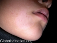
Facial eczema (courtesy Dr. McColl)
2/ Babies (6-12 months)
Rash often appears on extensors of elbows and knees (places that are easy to scratch as they crawl)
3/ Childhood phase (2-10 years)
Often develop a mainly flexural presentation: elbows, knees, wrists, ankles and neck
Can also affect peri-oral and peri-orbital areas
With chronic rubbing of skin can get lichenification (thick skin with exaggerated skin markings. Lichenification usually a bigger problem in darker skin types)
Oozing and crusting with secondary infection can occur
Flexural atopic dermatitis (globalskinatlas)
Flexural eczema (courtesy Dr. McColl)
Lichenification (courtesy Dr. McColl)
Adolescent phase (12-20) to adult phase (>20)
Atopic dermatitis often improves although barrier function is always decreased
Flexural eczema may still be present and eczema may be characterised by thick lichenified plaques from scratching
With severe eczema can get prurigo nodules as a reaction to chronic rubbing. These appear as nodules.
These can be extremely itchy and need strong topical steroid to penetrate this thick skin
Patients often start to develop more localised eczema (eg hand dermatitis or peri-ocular dermatitis) which may also be irritant or allergic (see contact dermatitis section for more info on this)
Hand dermatitis
Periocular dermatitis
Clues to diagnosis of atopy:
Atopic patients may have ichthyosis vulgaris (due to filaggrin mutatoin - discussed later) so may also have:
Hyper-linear palms of hands
Keratosis pilaris
Sub-types of eczema:
Discoid eczema:
Common type of eczema that can occur in atopic eczema
Get scattered, roundish patches of eczema that can be intensely itchy
Often need a potent topical steroid to get discoid eczema under control
Discoid eczema
Pityriasis alba:
Common manifestation of atopic eczema in younger people
Especially in darker skin type
Hypopigmented scaly areas seen: often on face but also on back and thighs (may be mistaken for pityriasis versicolor or even vitiligo)
Pityriasis alba
INFECTION
Having atopic skin puts you at increased risk of infection (including bacterial, viral, fungal and molluscum infection)
Bacterial infection:
If skin is particularly weepy or see honeycomb crusting think bacterial infection
Atopic dermatitis patients tend to be colonised with staph aureus (30-100%)
Colonisation increases the risk of infection
MRSA:
11-34% of AD patients colonised with S aureus have MRSA
MRSA associated with more severe eczema
If have an eczema patient who is very resistant to treatment you should consider swabbing skin to make sure they don’t have MRSA
MRSA carriage eradication example (but should look up local guidelines):
1/Mupirocin in nares tds for five consecutive days
2/An appropriate wash (eg octenisan) daily x 7 days
For more on microbiome and MRSA infection click on link
PVL (Panton valentine leuckocidin) staph aureus:
PVL is a toxin produced by certain types of S aureus
The toxin can kill white blood cells and cause damage to skin and deeper tissues
Clinically it can present with cellulitis, abscesses, boils and folliculitis so suspect in eczema patient who is getting recurrent boils
More about PVL at end of atopic eczema section
Streptococcal infection eczema:
Can also get Grp A streptococcal infection in atopic dermatitis patients
May see pustules with streptococcal infection and can be painful
Eczema herpeticum:
Dermatological emergency
Occurs when atopic eczema becomes secondarily infected by herpes virus
Suspect if get sudden flare up of eczema and child is unwell (fever, lymphadenopathy etc)
Look for small blisters and classical ‘punched out erosions’
If it is around the eye needs urgent ophthalmology review to outrule eye involvement
Important to recognise early and to put patient on oral or IV antivirals
[for dosing look at BNF and treat as for immunosuppressed patient as skin is ‘locally immunosuppressed’ in eczema]
Can use acyclovir
Famciclovir actually shows better bioavailability so may be more effective but it is more expensive
Often becomes secondarily infected with bacteria so look for crusting and treat with antibiotics
Infection in atopic skin with coxsackie virus (eczema cocksackium) and chickenpox can cause similar clinical picture

PATHOGENESIS
A complex range of factors at play:
Skin barrier dysfunction
Immune dysregulation
Environmental factors (allergens, altered skin microbiome)
Genetics
Their relative roles and whether they are all inter-related is still not fully understood
As it is so complex I will go through simplified versions but which will hopefully give a better understanding of the various current therapeutic approaches
Ways to conceptualise it:
Primary problem is a barrier defect which leads to immune dysregulation (outside in theory)
Primary problems is immune dysregulation which leads to the barrier defect (inside out theory)
Eczema as a barrier defect:
Keratinocytes in skin start out at basal layer and during life cycle work there way to the surface
At the surface they become flattened and are known as corneocytes
This outermost layer of the epidermis is called the stratum corneum
The stratum coreum provides the majority of the barrier function of skin
Think of the stratum corneum as a brick wall
The corneocytes are analogous to bricks
The lipid lamellae are analogous to cement
In eczema you have a barrier defect:
Therefore can’t keep water in skin as efficiently which can lead to dysfunction of the skin
Things from the environment get in to the skin like air pollutants, bacteria, irritants and allergens
These are presented to the dendritic cells of the immune system
These activated dendritic cells promote a shift from Th1 cell activity to Th2 cell activity
Increase Th2 cell activity leads to an increase in its signature cytokines such as IL 4, IL 5, IL 13, IL 31, IL 33
[IL-31 is a cytokine that produces itching]
With increased inflammation the patient itches and then scratches
This further disrupts the barrier and allows more antigens/bacteria in creating an itch-scratch cycle that makes the eczema worse
Th2 cytokines also:
Downregulate important proteins in skin (eg filaggrin)
Facilitate transcutaneous sensitisation
Facilitate bacterial binding and infection
As disease gets more chronic you get upregulation of other T helper cells and their assosciated cytokines:
Th17 (IL 17)
Th22 (IL-22)
Th1 (IFNy)
Agains this is a simplified version of the eczema pathway which involves environtmental factors further degrading the barrier, chemicals released from keratinocyts (eg TSLP), eosinophils and increased production of IgE that I will not go in to here
I will touch briefly on genetic factors below
Genetic factors and skin barrier dysfunction:
Multiple genetic factors have been identified that can lead to a defect in the skin barrier
Mutations in filaggrin are the most well documented (at least 20 loss of function causing filaggrin deficiency have been identified)
Lack of this protein in the skin causes an inherited dry skin condition called ichtyosis vulgaris and is strongly linked to development of atopic eczema
Filaggrin is vital for skin cells to mature properly into the tough, flat corneocytes that form part of the stratum corneum
Filaggrin is formed from the breakdown of profilaggrin which is a protein contained in the granules of the granular layer of the epidermis
After cleavage, liberated filaggrin binds to and collapses the keratin cytoskeleton, resulting in flattened keratin cells aligned parallel to the outer surface of the epidermis. This improves barrier function.
Therefore without filaggrin the keratinocytes are not aligned properly and antigens can easily enter and meet antigen presenting cells causing an immune response
With fillagrin intact
Filaggrin not functioning
Also, when filaggrin is degraded its degradation products are important in:
1/retaining moisture (natural mositurising factor)
2/killing bacteria
3/maintaining skin pH
Therefore not having enough filaggrin leads to impaired barrier function, inability to retain water, dry skin, abnormal pH and too much bacteria .
Filaggrin mutations are associated with:
Increased risk of atopic eczema
Increased risk of allergic sensitisation
Increased risk of peanut allergy
But not all cases of eczema are related to filaggrin issues:
70-80% of eczema cases worldwide have no filaggrin mutations
(FLG mutations found in 18% British cohorts)
Other genetic candidates are being explored which can cause impaired barrier function by causing:
Increased proteases in skin
Decreased protease inhibitors
Problems with adhesion proteins between corneocytes
SEVERITY ASSESSMENT
It is good to assess how much the eczema is affecting the patient
Should ask aout sleep disturbance, missed days of school/work
Quality of life questionnaires:
DLQI (how it affects your quality of life over the last weeks: scored out of 30)
POEM (Patient oreientated eczema measure - more specific to eczema. Again how it affects QoL over the last week. Scored out of 28)
Clinical severity assessment scores:
EASI
Looks at 4 body regions (head/neck, trunk, upper limbs, lower limbs)
Assesses the area affected in each region
Looks at redness, thickness (induration, papulation, oedema), scratching (excoriations) and licenification
Get combined score much like PASI
SCORAD is another severity assessment score sometimes used
MANAGEMENT
General:
Very important to take time particularly in the first consultation to counsel on what eczema is and why you use topical treatments
When counselling patients or parents can use the analogy of a house made with brick walls
The emollient is very important in keeping the brick walls well maintained and in good condition
However if the house catches on fire (skin is red) then topical steroids (or other anti-inflammatories) will be needed to put this fire out
CLEANSING
Soaps, shower gels, shampoos and baby bubble baths are very irritant and should be avoided
Soap substitute or emollient bath additive should be used instead. Can also use these to wash hair.
Fragrances and perfumes are also known irritants to skin and should not be used.
When bathing child- only use bath for 5-10 minutes. As soon as finished pat (instead of rubbing) down child with towel. Then use a greasy ointment such as emulsifying ointment as soon as they are out and this will lock in the moisturiser (‘soak and seal’)
Examples of soap substitutes
Dermol 500, QV gentle wash, hydromoll bath and shower, doublebase emollient wash gel
Bath additives examples:
Oilatum junior bath additive, doublebase bath additive, hydromol emollient bath additive, E45 emollient bath oil
EMOLLIENTS
Hydration is key in dealing with atopic patients
Use as much emollient as possible
When estimating how much emollient is enough to prescribe:
The average needed for 2 applications daily for 1 week is:
500g in adults
250g in children
When using emollients we ask parent to use a clean spoon when taking it out of the tub as if use hands when taking out emollients it may introduce bacteria into the tub
The greasier an emollient is the better it will hydrate the skin (eg Hydromoll ointment)
But it is often about finding the best emollient that parents will actually adhere to using as some parent find some ointments are far too greasy and stick on clothes
Ointments- have less ingredients so child less likely to react/develop allergy to an ointment. Therefore they are generally preferable to creams but they can be very greasy so sometimes as child gets older become less practical.
Creams- contain emulsifiers, preservatives and antimicrobials and may sting more than ointments.
Common emollients used in UK:
Ointments: Hydromol ointment, Epaderm ointment, Diprobase ointment, QV ointment, Cetraben ointment
Emulsifying ointment tends to be used in Ireland
Creams: Aveeno cream, diprobase cream, cetraben cream
Silcocks base tends to be used in Ireland
From light consistency to greasy consistency:
Aveeno, Zeroveen, Balneum, Doublebase gel
Zerobase
Cetraben cream
Emollin spray (50:50 spray)
Cetraben ointment
Zeroderm ointment
Hydromoll ointment
50:50 ointment
Parents often ask if child can go swimming.
In general it is okay to use swimming pools.
Tips would be to mositurise the baby before swimming.
Use a whole body swim suit as otherwise baby (helps with grip as otherwise baby would be slippy)
As soon as baby is out use a soap substitute if possible (to wash off the chlorine which can be irritating) and then moisturise afterwards
TOPICAL CORTICOSTEROIDS (TCS)
Extremely important in managing eczema
Often times parents have steroid phobia but if used correctly are very safe
Important to use appropriate strength of steroids depending on location of the rash and patients age
In general use weaker steroids in areas of thin skin (face and eyelids) and can use stronger steroids in areas of thick skin (palms and soles of feet) with skin on the body somewhere in between
Steroid potencies (commonly used bold)
Weak: Hydrocortisone 1%
Moderate: Eumovate (clobetasone butyrate), Betnovate rd (1/4 strength betnovate), Haelan tape (fludroxycortide)
Potent: Betnovate (betamethasone 0.1%), Elocon (mometasone), Synalar (fluocinolone)
Super potent: Dermovate (clobetasol propionate)
Combination topicals:
Fucidin H: hydrocortisone and fucidin (weak steroid, antibacterial)
Daktacort: hydrocortisone and clotrimazole (weak steroid, antifungal)
Betnovate C: betamethasone valerate and cliquinol (antibacterial): generally preferable to fucibet as resistance to fucidin is common
Fucibet: betamethasone valerate and fucidin (strong steroid, antibacterial)
Trimovate: Eumovate/neomycin/oxytetracycline (moderate steroid, anti-fungal, anti-bacterial)
Lotriderm: Betamethasone diproproionate and clotrimazole (strong steroid, anti-fungal)
Dermovate NN: Dermovate, neomycin, nystatin (super-potent steroid, anti-bacterial, anti-fungal)
Generally ointments are preferable to creams as there is less preservatives in them.
Steroids should work quickly, if child is not responding within a week then you might not be using enough steroids, steroid is not strong enough, or something else is wrong
One potential routine:
Many potential routines for topical steroid use exist, here is one such example:
When a flare is present: often give the appropriate steroid daily for 7-10 days (no major benefit from using twice daily with single steroid preparations)
Then when it is under control use it twice weekly as maintenance (using it twice weekly won’t cause any long term effects)
FINGER TIP UNITS
Finger tip units are a useful measure of how much steroid is needed to cover particular body parts
1 FTU is encough to treat an areas of skin twice the size of the flat of and adult’s hand with fingers together
In general:
Head to toe treatment for adults is 20-30g so daily treatment for 1 week 140g
Child 6-12 months: 5g so daily treatment 1 week is 35g
Child 4 years: 6g so daily treatment 1 week 42g
Child 6-10 years: 8g so daily treatment 1 week 56g
TOPICAL CALCINEURIN INHIBITORS (TCI)
Often used as steroid sparing agents
Particularly good for face and flexures
Might initially burn and sting but tends to get better with continued use
Should start using when skin not too badly inflammed to prevent stinging (so might want to give topical steroid for few days initially to get flare under control)
Pimecrolimus (elidel)- 1/3 as potent as tacrolimus
Tacrolimus (protopic)- comes in two strengths
0.03% licensed > 2 years
0.1% licensed > 16 years
(Stronger strength often used off license in younger age groups in dermatology)
Can do different routines for example:
Acute flares twice daily for 3-4 weeks
Maintenance: twice weekly up to 1 year (appears to reduce number of eczema flaress)
Or other types of routine such as topical steroid x 1 month, then protopic x 1 month
Side effects:
Increased HSV has been reported so ask them to stop if develop cold sores but in general in practice doesn’t appear to be major issue
Theoretical risk of increased risk of skin cancer as is an immunosupressant but this doesn’t seem to be the case after decades of clinical practice (black box warning so may want to warn patients about this in advance so they are not alarmed)
[APPLES study: no evidence found that topical tacrolimus increases long-term cancer risk in children with AD after 10 years follow up
Children must of used tacrolimus for 6 weeks]
NOVEL TOPICAL AGENTS
Crisabarole
PDE4 inhibitor - ‘Like topical apremilast’
Licensed in USA since 2016: moderate effect ‘at best’
Recent FDA approval (December 2020): infants > 3 months
Ruloxitinib
JAKi
Recent phase 2 study shows superior to mid-strength steroids (Kin et all 200, JAACI)
Increasing strength of of ruloxitnib get increasing response in EASI scores
Some reports here of 80% improvement in EASI in sites used
True AD-1 and True AD-2 studies sample 1250 patients and in essence replicated the above study
OCCLUSION AND GARMENTS
Occlusion can be beneficial by improving hydration, steroid penetration, acting as a barrier to scratching and improving sleep
Methods:
Paste bandages
Wet wraps
Garments
Occlusive dressings
Good education is needed on the use of occlusion
The BAD website: skinhealthinfo.org.uk has good informational videos on how to apply occlusive dressings, paste bandages and wet wraps (supports and resources: video guides)
Occlusion cautions:
Increases potency of steroid used so use steroids in appropriate areas (not face, flexures)
Can exacerbate active skin infection
Paste bandages:
Single use only
Icthopaste (6.32% zinc oxide), Viscopaste (10% zinc oxide), Zipzoc bandages (20% zinc oxide)
Icthopaste and viscopaste are cotton wrap around bandages
Zipzocs are cotton stockings (often easier to apply than wrap around bandages above but children are too small to use them)
They have soothing and anti-inflammatory pathways and can improve penetration of topical steroids due to occlusion
You need to cover bandages and stocking with a tubular dressing to avoid soiling of clothes and to help keep them in place
A tip is to put the paste bandages in the fridge before application which can be soothing
Products can be left in place for seven days if indicated but obviously often changed more frequently depending on need for emollient and topical steroid
Wet wraps:
Most commonly used with emollient underneath but can be used with topical steroids underneath for short periods of time
When applying will need garments: 2 tops, 2 leggings, warm water and emollient cream (to cover smaller areas can use tubular bandaging instead)
After washing you apply a thick layer of cream
Wraps are prepared by putting 1 top and 1 leggings in a bowl of warm water, then squeezing garments of excess water so they are damp but not dripping wet
They are then applied to the patient (make sure labels/seam are on the outside) and then put dry top and leggings on top
Can be left on for 24 hours but often are left on overnight and taken off in the morning
Caution:
If think have skin infection do not use them as can make them worse
Can be uncomfortable for patients particularly in cold weathere
Eczema garments:
Are sometimes referred to as ‘eczema clothing’
They are desinged for individuals with eczema and other similar conditions
They aim to reduce itching and prevent scratching
For babies they may include mittens and footies and may have a head garment
Types of garments from cheapest to most expensive:
Clinifast (viscose)
Skinnes (viscose)
Skinnies (silk)
Dreamskin
Occlusive dressings:
Hydrocolloid dressings that are applied over treatment to improve absorption of the medication and prevent damage from scratching
It increases the strength of the steroid so should be used only for a short period of time and should not be used if the skin is affected
Avoid occlusive dressings on face or flexural areas
An example of an occlusive dressing that can be used is duoderm extrathin
BLEACH BATHS
Can be used with the proposed aim to reduce bacterial load (eg S Aureus) and as such to reduce Eczema severity so may consider if patients get recurrent secondary skin infections
How to use:
Use pure bleach without fragrance or soap
Examples:
Tesco everyday value thin bleach
Milton disinfecting solution
ASDA Smart Price thin bleach
Run bath with no additives:
Add half a cup (150ml) of bleach to at least 10cm depth of water in an adult bath
(For baby tub use 2ml bleach for each 1L water)
Mix well
Soak for 10-15 minutes
Can use an emollient cream to clean skin when soaking
Can wet face and hair (should not cause harm if gets in eyes or mouth) but make sure child is not drinking it
Then rinse with plain water, pat skin dry and apply emollients as usual
Use once or twice a week
Telling parents to use a bleach bath can sometimes alarm them so can use the analogy that it essentially is like ‘making your own swimming pool‘
PHOTOTHERAPY
Narrowband UVB:
Time consuming but can be useful for management of eczema
Not as effective as for psoriasis and may need longer treatments
Important to emphasize frequent moisturising as it is good for inflammation but the skin can dry out
CONVENTIONAL SYSTEMIC AGENTS
In general Methotrexate and Ciclosporin are the commonest conventional systemic agents used
Ciclosporin - works well and quick so is a good rescue drug to break the cycle of eczema and get it under control. But time and dose limited. With long use get kidney problems and hypertension
Azahioprine and Mycophenolate mofetil are other options but tend to be used less commonly then the above agents and due to the advent of new biologic agents
Azathioprine appears to have similar efficacy to methotrexate but get increased risk of skin cancers
In general should try to avoid oral steroids in atopic eczema. May work at the start but when they are stopped may get worsening flare of condition.
TREAT trial:
A recently published large, multi-centre RCT
Compared Methotrexate (0.4mg/kg/week) and Ciclosporin (4mg/kg/day) in a paediatric population (aged 2-16)
Findings seem to suggest that ciclosporin works quicker in decreases eczema severity (at week 12) but from about week 20 onwards the picture starts to flip and methotrexate seem to be better
In study both drugs were quite well tolerated and appeared to be safe
Also in the study methotrexate was commenced straight away at the full treatment dose (0.4mg/kg/week) instead of starting at a lower dose and titrating upwards and this appeared to be well tolerated
Here are links to the releavant medications (in systemic part of website):
ANTI-HISTAMINES
Don’t really work well for itch of eczema!!!
Occasionally sedating anti histamines can be used if patient not sleeping but should only prescribe very short course of this
TARGETING CYTOKINES
DUPILUMAB
Dupilumab was the first targeted biologic treatment approved for atopic eczema
Fully human monoclonal antibody to an IL-4 receptor subunit
Targets IL 4 and IL 13 (TH2 cytokines which are increased in eczematous skin)
Both receptors for these cytokines contain the same subunit IL-4RA
Dupilumab targets this subunit
IL-4 and IL-13 are both cytokines produced by TH2 cells
Increased IL-4 and IL-13:
Impairs keratinocyte differentiation
Lower anti-microbial peptide production
Increases eosinophil recruitment
Causes further TH2 differentiation and survival
PHASE 3 trials: SOLO 1 and 2
Approximately 50% of patients reached an EASI 75 at 16 weeks
Dupilumab EU approval:
October 2017 adults
November 2020 children 6-11
Indication:
Moderate to severe eczema who have failed at least 1 systemic agent
Other indications (outside dermatology):
Chronic rhinosinusitis with nasal polyposis
Severe asthma failing to respond to conventional treatments
(if patients have asthma and are given dupilumab for eczema you need to make them aware that the asthma could rebound if it is discontinued)
Dosing in eczema:
Adults: 600mg subcut induction, then 300mg every 2 weeks (weight > 50kg)
Adolescent (12-18 year olds)/adults < 50kg: 400mg induction, then 200mg every 2 weeks
Review:
At 16 weeks
Continue treatment if adequate response (ie 50% reduction in EASI or 4 point drop DLQI)
Potential protocol:
Document prior treatments
Record baseline DLQI, POEM, EASI
Routine bloods and pre-immunosuppression screen (no need for CXR/Quantiferon)
Contraception for females (can’t know for sure it is safe in pregnancy. Has 11-week washout period)
Consider prophylactic hyloforte (lubricating) eye drops four times a day for 2 weeks prior to starting treatment (can increase to 8 times/day if symptomatic)
Potential monitoring:
Review at 8-weeks: enquire about eye symptoms, ? facial worsening, initial efficacy
Review at 16-weeks: Routine bloods, DLQI, EASI, POEM and decide whether to continue
Review 6 monthly thereafter: routine bloods, DLQI, EASI, POEM
Side effects:
Dupilumab induced ocular surface disease:
Commonest side effect
Incidence 30-50%
Occurs early
Often will require opthalmology review
Could consider giving hyloforte 4 times/day prophylactically and increasing to 8 times/day if symptomatic
Opthalmology team could consider steroid or ciclosporin drops if appropriate
Some patients need to stop dupilumab and in some cases can get scarring which is permanent
Dupliumab facial redness:
Can sometimes occur (may be more likely to occur in people who have facial eczema already or eye disease)
In these cases can consider reducing the frequency of dupilumab to 4 weekly
Other treatments that could be considered would include itraconazole orally, protopic or topical steroid but they are not always effective
Upper respiratory tract infection
In some patients: exacerbations of eczema and asthma
Eosinophilia
Dupilumab induced arthritis:
5% enthesitis (affecting elbows and achilles)
Can get arthritis of small and large joints
To manage reduce frequency of dupilumab
Can consider giving concomitant Methotrexate
Not considered to be too immunosuppressant (eg no increased risk of TB or opportunistic infections)
TRALOKINUMAB
Monoclonal antibody targeting IL 13 cytokine itself
Phase 3 clinical trial:
ECZTRA 1 and 2
EASI 75 33% at week 16
(compared less favourably to dupilumab studies EASI 75 45-50%)
When topical steroids allowed: EASI 75 38.9% at week 16
LEBRIKIZUMAB
Another anti-IL 13 agent
Phase 2 clinical trial (TREBLE)
EASI 75 60% at week 16
All groups used twice daily topical corticosteroids
So from this phase 2 trial appears that it is better than tralokinumab
The anti-IL 13 agents don’t seem to cause as many eye issues compared to dupilumab so the eye issues appear to be an issue with targeting IL-4
NEMOLIZUMAB
Humanised anti IL-31Rα monoclonal antibody
So stops the function of IL-31 which can drive itch
It is currently being trialled in eczema and nodular prurigo
Issues:
Can make asthma worse so need to be very cautious in asthmatic patients
URTIs
Nasopharyngitis
Clinical trial:
Unusual parameters used compared to usual trials:
Endpoint assessed at week 24
EASI score reduction 68.8%
JAK INHIBITORS
MECHANISM OF ACTION
Enzymes that add phosphate groups are called protein kinases
The Janus kinase (JAK) protein family is a family of kinases that transduces signals via the JAK-STAT pathway
There are 4 members in this family
The JAK-STAT pathway is a signalling pathway that can mediate cellular responses to a variety of cytokines
This pathway is used by T cells
Different T helper cell subsets (eg Th2, Th1, Th 17) can have different JAK-stat receptors
How the pathway works:
A cytokine binds to the receptor and as a result JAK adds a phosphate to the opposite side of the receptor (cross phosphorylation)
2. This attracts the STAT protein which docks to the receptor
STAT is phosphorylated again and they then bind to each other forming a dimer
The dimer then moves into the nucleus, binds to the DNA, and initiates the transcription of genes
Different cytokines use different JAK family members to signal
JAK 1 is common in cytokine signals in eczema so all agents target JAK 1
No JAK inhibitors are entirely specific or targeted
Some JAK inhibitors are not specific at all and some are more specific
JAK inhibitors used in eczema:
Can be considered selective (ie focus on JAK 1) or non-selective (ie focus on JAK 1 and 2)
All are oral tablets taken once daily
Two doses are given for each agent which gives more flexibility to use
Half life is very short so can have tight control (by 24-48 hours is mostly out of system)
Eczema tends to come back quickly if stopped
Stop if don’t have adequate response by 16 weeks
Primarily renally cleared
Baricitnib: Targets JAK 1 and 2
Abrocitinib: Targets JAK 1
Upadacitinib: Targets JAK 1
Ruloxitinib (cream): Targets JAK 1 and 2
In other conditions such as alopecia areata the JAK inhibitors may also be selective for JAK 3
JAK signalling is key to multiple processes including:
Inflammation (having effects on T cell/B cell proliferation and function)
Anti-microbial response: anti-viral, anti-mycobacterial
Haematopoeisis
Anabolic metabolism
JAK 1 signalling is involved in:
Inflammation
Anti-viral activity
Anti-tumour activity
So if block JAK 1 activity:
Get increased risk of viral infection (eg herpes simplex/herpes zoster)
Also may get decreased tumour surveillance
JAK 2 signalling is involved in:
Granulopoiesis
So if block JAK 2 activity:
Get increased risk of neutropaenia and granulpaenia
ADVERSE EFFECTS JAK INHIBITORS
Very common (>1/10)
Nausea
Common (1/10 to 1/100)
Herpes simplex, herpes zoster
Headache, dizziness
Vomiting, upper abdominal pain
Acne
CPK elevations > 5 times ULN
Uncommon (1/100 to 1/1000)
Pneumonia
Thrombocytopaenia, lymphopaenia
Hyperlipidaemia
Diverticulitis, bowel perforation
Venous thromotic events (DVT, PE, some reports of sinous venous thromboembolism)
Need to ask about personal and family history of blood clots
Numerous systematic revie and meta-analysis don’t seem to suggest DVT/PEs are increased with JAKi but was seen in trials with tofacitinib so FDA has put a warning in
There is also a signal that JAK inhibitors could potentially increase risk of malignancy so would need to be very cautious in person with history of malignancy (ideally would use biologics targeting cytokines instead)
Some side effects are more class specific:
Abrocitinib: increased risk thrombocytopaenia and nausea
Upadacitinib: increased risk acne, anaemia/neutropaenia
Baratinib: Risk of diverticulitis and bowel perforation being monitored. If there is a linke appears to be rare
SCREENING AND MONITORING
History/exam:
Consider risk factors: HSV, VZV, DVT, PE, Clotting family histor, CV risk factors or disease, smoking
Invesigations:
FBC
U&E
LFT
Lipids
CK
Hepatitis/HIV serology
TB screen: CXR/Quantiferon
Potential monitoring (with bloods at each appointment):
Nurse review 4 weeks (with bloods, particularly FBC)
Then review at 4 months - decide if to continue
Then 4 monthly x 1 year
Then 6 monthly
BARACITINIB
Targets JAK 1 and 2
Dose 2mg/4mg
Half life: 12.5 hours
4mg is best dose
BREEZE AD-1:
EASI 75 @ 16 weeks 24.8%
Adverse effects: 56% at 4mg
Serious adverse effects: 1.2%
BREEZE AD-4:
Combined Baricitinib/Topical corticosteroid vs Placebo/Topical corticosteroid
EASI 75 @ 16 weeks:
45% Baractinib Vs 20% Placebo
As targets JAK 2 also there is an increased risk of anaemia/thrombocytopaenia
ABROCITINIB
Targets JAK 1
100mg/200mg
Half life: 5 hours
JADE study:
EASI 75 response rate 62.7% at week 12
EASI 90 response: 38.6%
200mg will be the licensed dose adults
100mg (in adolescents and people > 60 and people with decreased renal/hepatic function)
Works very quickly
JADE compare:
Abrocitinib vs dupliumab (all in combination with topical therapy)
Abro 200mg best: EASI 75 70.3%
Abro 100mg: EASI 75 58.7%
Dupilumab: EASI 75 58.1%
Placebo: 27.1%
UPADACITINIB
Targets JAK 1
15mg/30mg
Half life: 8-14 hours
30mg adults
15mg adolescents
Will be licensed for people from 12 years of age
At 16 weeks:
EASI 75 of 72-79%
EASI 90 also very good
RCT comparing Upadacitinib vs Dupilumab
(Blauvelt et al, JAMA Dermatol, 2021)
N=924
EASI 75 week 16: Upadacitinib 71% vs Dupilumab 61%
But there was increased risk of significant infection in upadacitinib group
MONOTHERAPY TRIAL RESULTS SUMMARY
Lack of comparison trials currently so can’t make definitive conclusions as different studies had different methods etc
Below is results of EASI 75 at 12-16 weeks for the big monotherapy clinical trials for each agent
MISCELLANEOUS TOPICS:
ECZEMA, BACTERIA AND MRSA
The skin microbiome is complex and diverse
There is a wide variety of microrganisms (including fungi, viruses and bacteria)
It differs from anatomic site to anatomic site and this can be seen as early as day 2 in life
(eg infants delivered vaginally have microbiome rich in lactobacilli, if born by CS it is initially riched in organisms colonising mother’s skin)
The microbiome changes over time in response to may things (eg pets, rural environment)
The microbiome is very diverse in normal skin and also diverse in quiescent atopic skin.
Eczema disease severity correlates with reduced diversity and Staph aureus colonisatoin
Atopic dermatitis patients tend to be colonised with staph aureus more often then control population
Staph aureus colonised 30-100% of atopic dermatiits patients
Lesional skin > non-lesional skin and nose
Colonisation:
Increases risk of infection
S aureus toxins can act as superantigens activating lymphocytes and macrophages
S aureus can trigger multiple other aspects of the immune system, eg:
Initiating prodcution of TSLP which triggers a switch to Th2 skewing
S aureus may displace normal flora of skin (eg s epidermis)
Normal flora produce substances that decrease inflammation.
So overpopulating skin with pathogenic staph you lose a mechanism to suppress inflammation
MRSA:
11-34% of AD patients colonised with S aureus have MRSA
MRSA colonisation is associated with:
Eczema severity
Hospitalisatoin
Use of TCI and TCS
Having a healthcare worker at hom
Oral antibiotic therapy
If have an eczema patient who is very resistant to treatment you can swab skin to make sure they don’t have MRSA
MRSA carriage eradication:
1/Mupirocin in nares tds for five consecutive days
2/An appropriate wash (eg octenisan) daily x 7 days
To treat or not to treat Staph aureus colonisation:
For:
Colinisation with toxin secreating S aureus can contriute to pruritus and inflammation
Coloinsation is a risk for infection
Against:
S aureus is a common commensal
Eradication is difficult
Cochrane systematic review showed no clear evidence of benefit (although studies were small)
PVL TOXIN
PVL (Panton valentine leuckocidin) staph aureus:
PVL is a toxin produced by certain types of S aureus
The toxin can kill white blood cells and cause damage to skin and deeper tissues
Clinically it can present with cellulitis, abscesses, boils and folliculitis so suspect in eczema patient who is getting recurrent boils
Risk factors for PVL (5 Cs):
Close contact (eg contact sports such as rugby)
Cuts and grazes
Contaminated items (eg touching contaminated gym equipment, towels, razors)
Crowding (eg living in military accomodation, prisons, boarding schools)
Cleanliness (an unclean environment encourages the bacteria to spread)
Clinically it can present with cellulitis, abscesses, boils and folliculitis so suspect in eczema patient who is getting recurrent boils
To diagnose take a standard bacterial swab and make sure you state on the form that you would like them to analyse for PVL toxin otherwise they won’t look for it
Management:
PVL carriage should be treated with octenisan wash and nasal ointments similar to regimen for MRSA decolonisation
If have active skin and soft tissue infection look up local guidelines and/or discuss with microbiology:
For instance:
Abscesses can be incised and drained
If PVL + but not MRSA: Some regimens suggest Flucloxacillin
If PVL + and MRSA +ve: Some regimens suggest rifampicin and another antibiotic
Severe infections may require IV antibiotics
PVL related pneumonia can be a complication