For this part of the website I will go through very basic schematics of the skin. I will go into more detail of various structures at the relevant sites. This section aims to outline the different layers of the skin. The basic structures within it and the cells that might be encountered within the skin.
The skin is composed of:
1/ An outer epithelial layer- epidermis
2/ Underlying connective tissue layer- dermis
The dermis lies on the subcutaneous tissue which is composed predominantly of adipose tissue
Hair follicles and other adnexal structures actually often extend down to subcutis despite this illustration.
Epidermis:
Very simplistic schmeatic of keratinocytes in epidermis
The predominant cell in the epidermis is the keratinocyte
The keratinocyte makes its way from the basal layer up towards the surface during its life cycle. During this time it changes its composition and gets flatter
The keratinocytes are arranged in layers
1/Basal layer is the deepest layer. The basal cells are the epidermal stem cells and divide.
50% of the daughters of stem cells remain as stem cells while the other 50% leave the basal layer and enter the spinous layer.
2/ Prickle cell layer (stratum spinosum)
Can vary in thickness depending on location
Called prickle layer as the desmosomes which connect the cells of the epidermis are very evident in this layer on H&E staitning
3/Granular layer (stratum granulosum)
Contains numerous keratohyalin granules which contain a protein important in eczema called filaggrin (see eczema section) among other substances
Looks blue on histology
4/Clear cell layer (stratum lucidum)
Stains poorly. Only seen in thick skin.
5/Horny layer (stratum corneum)
The outermost layer. It contains corneocytes which are keratinocytes that have completed their differentiation program and have lost their nucleus and cytoplasmic organelles. These cells are constantly shed from the epidermis.
This layer provides the vast majority of the barrier function of the skin.
Think of the stratum corneum as ‘brick and mortar’
The bricks consist of corneocytes.
The mortar consists of a variety of lipids and other substances surrounding the corneocytes which are also important in barrier function.
Thickness of epidermis varies from thin in eyelids (0.1mm) to thick in palms and soles (1mm)
Keratinocytes are held together by proteins called desmosomes which are made up of different subtypes of proteins called desmogleins.
If certain desmogleins are missing/attacked you get superficial blistering which often presents as erosions.
Under the epidermis and connecting the basal keratinocytes to the basement membrane are the hemidesmosome proteins.
The basement membrane is a complicated structure and there are a variety of proteins involved in joining the epidermis to the dermis.
The basement membrane is important in relation to blistering diseases.
Below is a schematic of the Basement membrane zone (BMZ) that shows some of the proteins involved in the area (even this is a simplistic version)
Blisters due to missing/attacked proteins in the BM tend to be more tense compared to blisters due to splitting higher up in the epidermis
In general, if the split occurs above or within the lamina lucida there will be no scarring.
If split occurs below the lamina lucida there will be scarring.
Other important cells in epidermis:
1/Langherans cells:
Derived from Bone marrow
Dendritic cells that are members of monocyte/macrophage series of cells
These cells are antigen presenting cells- they envelope antigens that penetrate skin and take them to local lymph nodes where they present them to T cells to activate the adaptive immune response
2/Melanocytes
Derived from embryonic neural crest
Are found along the basal layer
In normal skin are found in about a 1:10 ratio to keraitnocytes along the basal layer
Have dendritic processes which provide pigment to overlying keratinocytes
They can cluster to form nests of benign looking nevus cells in benign nevi
If become malignant they form melanoma (cells will be atypical)
3/Merkel cell
Rarest cell in epidermis
Neural crest origin
Plays a part in sensation at nerve endings
If becomes malignant get a merkel cell carcinoma which has a very poor prognosis
Dermis:
Connective tissue of dermis consists of collagen and elastic fibres embedded in a ground substance
Collagen and elastic fibres are formed by fibroblasts
Upper part of dermis is called the papillary dermis
Lower part of dermis is the reticular dermis
The ‘ground substances’ between collagen and elastin fibres are glycosaminoglycans called hyaluronic acid and dermatan sulfate
[hyaluronic acid often is the material used as fillers in cosmetic derm to pump up lips etc]
Appendigeal structures in the dermis:
1/Hair follicles and arrector pili muscle attached
The epidermis itself extends into the hair follicle. The area from the epidermis to the sebaceous gland duct is called the infundibulum. As such, tumour of the epidermis can often spread down the hair follicle.
2/Sebaceous glands which can be found surrounding the upper part of the hair follicle. The sebaeous duct opens into the hair follicle and releases sebum into it.
3/Eccrine sweat glands open onto the skin surface and are found in most locations of the body
4/Apocrine sweat glands open into the hair follicles (above the sebacous duct) and are larger then eccrine glands. Are found only in certain locations body such as axilla, areola, genital areas, perianal area. The secretions the apocrine gland release are very odorous.
Dermal nerves and vasculature (blood vessels/lymphatics) are found in the dermis:
Get superficial dermal plexus and deep dermal vascular plexus.
Vessels also concentrated around the appendigeal structures.
Smaller blood vessels are more superficial.
Small blood vessels extend high up into the dermal papillae.
The epidermis is an avascular structure.
The skin encounters a host of toxins, pathogenic organisms and physical stresses. Therefore in addition to functioning as a physical barrier it can also be considered an active immune organ.
Immunological cells in skin play a very large part in many skin diseases. Immunological cells that may be found in the dermis include:
Neutrophil granulocytes
Eosinophil granulocytes
Lymphocytes (B and T cells-indistinguishable on light microscopy)
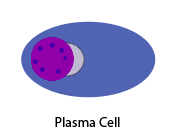
Plasma cells
Monocytes/macrophages
Mast cells
Mast cell


